The Ligament You’ve Probably Never Heard Of
Dec 07, 2022The Cutest Little Ligaments
Between the skin-we-touch and the muscles-we-palpate, lies a whole world of tissue and activity that is often under-addressed in our anatomy books, including one extraordinary structure we massage every day called the skin ligaments(Latin: retinacula cutis). They are made of connective tissue and part of the fascial system, but unlike most of the other ligaments you know, they do not connect bone to bone. These small, fibrous structures create a bridge connecting the skin’s dermis to the deep fascia – and you move them with every single massage stroke.
Where do they LIVE?
Skin ligaments can be found everywhere within the body’s cushion-y outermost layer, the subcutis, also called the subcutaneous tissue or superficial fascia. The subcutis is the dynamic space situated between the dermis and the deep fascia which stores energy, regulates temperature, exchanges hormones, and circulates lymph. In order for this busy community of cells and fluids to do their jobs, they require a stable structural network to live in, which is formed by none other than the skin ligaments.
What do they LOOK like?
The architecture of the subcutis is immediately visible in the anatomy lab once we go beneath the skin. Our eyes are first drawn to its most striking feature: billowy, bright-yellow fat lobules.
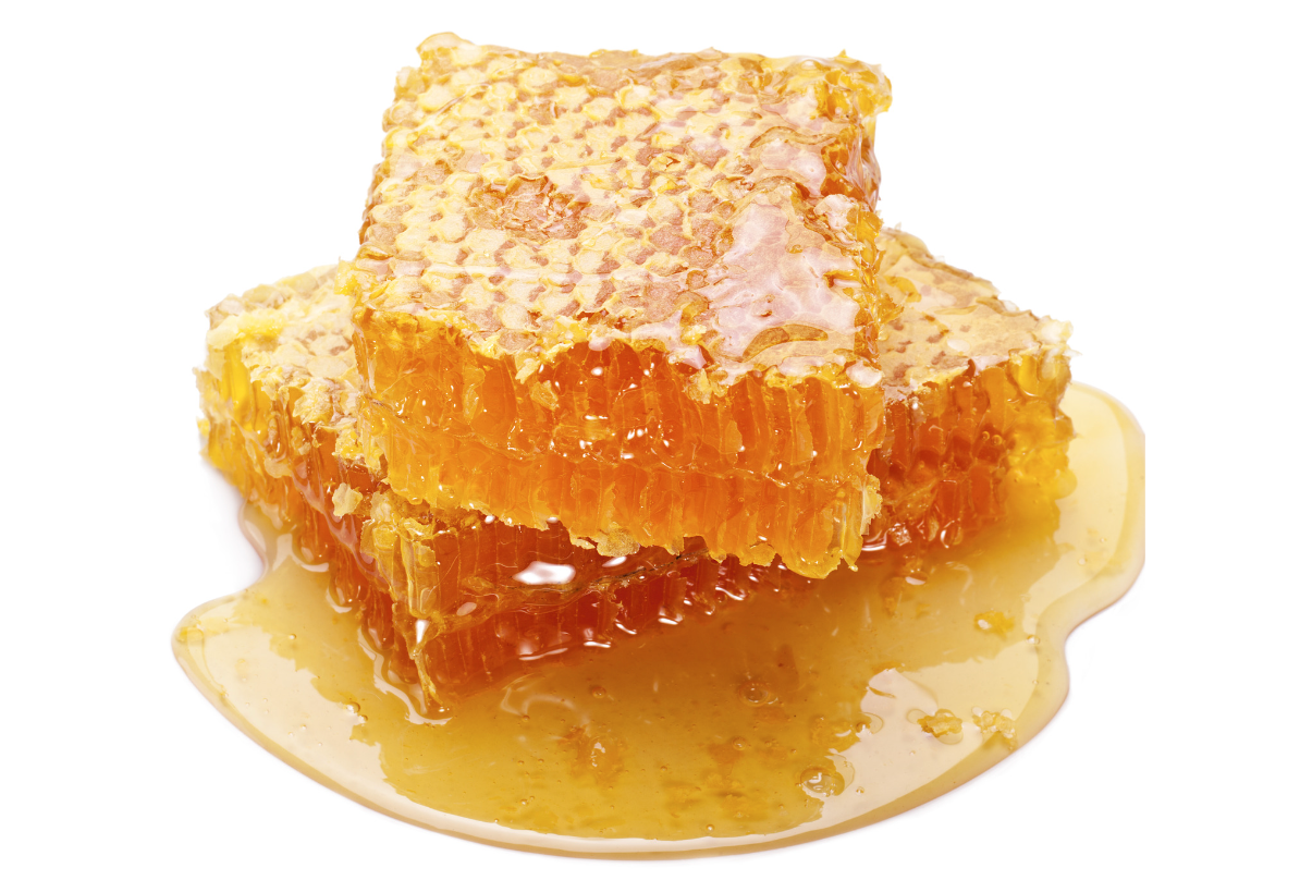
Looking a little closer we can see skin ligaments extending down from the dermis, between the fat lobules, d efining their shape and forming a fascial framework that resembles honeycomb or bubble wrap.
You have probably actually seen skin ligaments before, even if you haven’t been in an anatomy lab. Have you ever seen cellulite? As much as our culture may lead us to believe cellulite is a pathology, it is nothing more than the visible impression of your skin ligaments and fat lobules on the skin's surface.
What do they FEEL like?
Skin ligaments vary in thickness and density regionally and give structural boundaries to the fatty lobules. Areas that require more stability have shorter, denser, more tightly-packed-together skin ligaments, such as on the palms, soles of feet, and sacrum. In these areas the ligaments and lobules can feel like small, squishy, tapioca pearls (1mm-2mm). Areas that need to accommodate more internal or external movement between the skin and deep fascia have longer, not-so-tightly-packed skin ligaments such as on the abdomen and thighs. Here their adaptability makes them less easy to palpate, but you may perceive their texture similar to large boba or mini-marshmallows (5mm-20mm).
What do they DO?
Skin ligaments serve a number of functions which we would struggle to live without. Their containment of fat lobules provides a dynamic cushioning system that protects, insulates and disperses forces. The dividing walls in their “honeycomb” organization also protect nerves and blood vessels as they travel to the skin’s surface and prevent the fat lobules from just floating around. The morphological variation of their presentation – density and thickness – in different regions gives them the capacity to allow for dynamic movement or resist mechanical loading where needed. Vital to the functions of the subcutis, they also permit passage of lymph fluids. In short, the cute little skin ligaments serve the many needs of your most superficial layer in relation to the complex structure that is you!
Why we CARE.
As massage therapists, every time we move the skin we are also dragging the skin ligaments along for the ride. Understanding how they mechanically link the skin to the deeper body gives us a glimpse into the mechanisms at play in our most common massage techniques, particularly those that rely on shearing motions. From effleurage to petrissage, we use shearing forces all the time in massage.
We influence so much tissue before ever reaching muscle depth, understanding the anatomy of the skin ligaments can help us be even more specific with our touch and gives us a deeper appreciation for what lies just beneath the surface!
*If you like what you read and want to read more content like this, head over to Associated Bodywork & Massage Professional's website to read their latest issues of Massage & Bodywork magazine.*
REFERENCES & FURTHER READING:
For more information on the skin ligaments and the three-dimensional fascial anatomy of the subcutis, check out these resources:
Cotofana, & Kaminer, M. S. (2022). Anatomic update on the 3‐dimensionality of the subdermal septum and its relevance for the pathophysiology of cellulite. Journal of Cosmetic Dermatology, 21(8), 3232–3239. https://doi.org/10.1111/jocd.15087
Nash, Phillips, M. N., Nicholson, H., Barnett, R., & Zhang, M. (2004). Skin ligaments: regional distribution and variation in morphology. Clinical Anatomy (New York, N.Y.), 17(4), 287–293. https://doi.org/10.1002/ca.10203 In this foundational research, Nash documents the existence of the skin ligaments and their differing composition over the entire human body.
Stecco, Carla. (2015). Functional Atlas of the Human Fascial System. Churchill Livingstone Elsevier (Edinburgh). For a visual guide to the superficial fascia and skin ligaments, turn to Carla Stecco’s Functional Atlas of the Human Fascial System. She provides an extensively researched and visually documented tour of the subcutaneous region. Stecco is one of the world’s leading researchers on fascia, including the fascia of the subcutis region.
Sakata, Abe, K., Mizukoshi, K., Gomi, T., & Okuda, I. (2018). Relationship between the retinacula cutis and sagging facial skin. Skin Research and Technology, 24(1), 93–98. https://doi.org/10.1111/srt.12395
Wong, Geyer, S., Weninger, W., Guimberteau, J.-C., & Wong, J. K. (2016). The dynamic anatomy and patterning of skin. Experimental Dermatology, 25(2), 92–98. https://doi.org/10.1111/exd.12832
Are you curious about the mechanical link between the surface and deeper layers? The following research articles help establish the growing evidence that deformation of tissue at the skin level affects much deeper layers.
Engell, Triano, J. J., Fox, J. R., Langevin, H. M., & Konofagou, E. E. (2016). Differential displacement of soft-tissue layers from manual therapy loading. Clinical Biomechanics (Bristol), 33, 66–72. https://doi.org/10.1016/j.clinbiomech.2016.02.011
Pamuk, & Yucesoy, C. A. (2015). MRI analyses show that kinesio taping affects much more than just the targeted superficial tissues and causes heterogeneous deformations within the whole limb. Journal of Biomechanics, 48(16), 4262–4270. https://doi.org/10.1016/j.jbiomech.2015.10.036
Wilke, J., & Tenberg, S. (2020). Semimembranosus muscle displacement is associated with movement of the superficial fascia: An in vivo ultrasound investigation. Journal of anatomy, 237(6), 1026–1031. https://doi.org/10.1111/joa.13283


